GEFÄSSBIOLOGIE
Forschung und Lehre unserer Teams verbindet moderne Tierbiologie und Entwicklungsbiologie, aber auch medizinischen Anwendungen im Bereich der Gefäßbiologie. Diese umfassen sowohl Grundlagenforschung als auch angewandte Forschung. Die Forschungsschwerpunkte unseres Teams stellen sich wie folgt dar: Der Fokus in der Gefäßbiologie liegt in der Grundlagenforschung auf der Untersuchung der Blutgefäßentwicklung und -regression am Modell des Südafrikanischen Krallenfrosches Xenopus leavis. In der angewandten biomedizinischen Forschung liegt der Fokus auf Untersuchungen des Vasa vasorums der großen humanen Beinvene, relevant für die Herz-Bypass Chirurgie, Untersuchung von erworbenen und angeborenen Herzmuskelerkrankungen, Untersuchung der Tumorvaskularisation, sowie auf Studien über die Cannabinoid- (THC / THCV) Wirkung auf das Reproduktionssystem und auf die durch Diabetes mellitus (DM-2) induzierte, pathologisch veränderte Mikrovaskularisation ausgewählter Organe (Nieren, Leber, Herz, Auge etc.).
Des Weiteren befasst sich unsere Gruppe mit der Erforschung von NETs (neutrophil extracellular traps) im Kontext zu Parodontitis.Unsere Gruppe ist ein der weltweit führendes Kompetenzzentren auf dem Gebiet der Gefäßausgusspräparation (Vascular corrosion casting). Neben der Gefäßausgusstechnik betreiben wir Raster- und Transmissionselektronenmikroskopie (REM / TEM), 3D- Morphometrie, Histologie, und Intravitalmikroskopie. Kooperationen bestehen mit internationalen Forschungseinrichtungen und Universitäten, biomedizinischer Industrie, sowie mit Kliniken der Paracelsus Medizinischen Privatuniversität Salzburg (PMU / SALK).

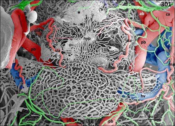
Pituitary gland. Vascular anatomy. Ventral view at the hypothalamo-hypophysial area. Shown is the arterial supply of the pituitary gland via terminal branches of the superficial infundibular artery (sia) and its drainage via the hypohysial vein (hv). Meningeal capillaries are colored green. ca cerebral carotid artery, dia deep infundibular artery, dl distal lobe of pituitary gland, hyp hypothalamus, il intermediate lobe of pituitary gland, me median eminence, nl neural lobe of pituitary gland, ab anterior branch of cerebral carotid artery, pb posterior branch of cerebral carotid artery. Asterisk marks large evasate.

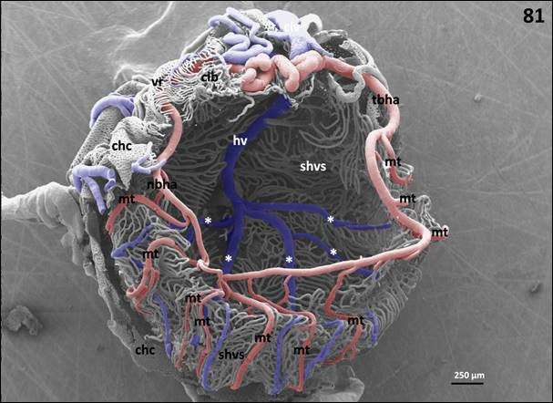
Superficial hyaloid vascular system of a left eye. Slightly oblique frontal view. Nasal is at left, temporal is at right. Two thirds of the chorio-capillaris (chc) and ciliary body (cib) are removed to expose the superficial hyaloid vascular system (shvs). Specimen is spatially distorted, but otherwise intact. Shown are nasal (nb) and temporal branch (tb) of the hyaloid artery (ha). Each semicircular branch gives off 7-10 meridional twigs (mt) which run towards the posterior pole of the eye. Further seen are the roots of the hyaloid vein (asterisks) between arterial meridional twigs (mt). hv hyaloid vein.
Fotos: Gefäßausgusspräparation / Vascular corrosion Casting (Quelle: COLOR ATLAS OF THE MICROVASCULATURE OF TISSUES AND ORGANS IN ADULT XENOPUS LAEVIS, A. Lametschwandtner und B. Minnich, in. prep.)


