VASCULAR BIOLOGY UNIT
The scope of our team is to connect modern animal biology and developmental biology as well as medical biology in the field of vascular research. This covers both, fundamental- and applied research. The vascular foci cover basic research on blood vessel development and regression in the African Clawed Toad, Xenopus leavis as model organism and biomedical research on coronary bypass surgery (Vasa vasorum of the Vena saphena magna), on acquired and innate disease of the cardiac muscle, on tumorvascularization and on cannabinoid (THC / THCV) effects on the reproductive system and on diabetes mellitus (type 2) induced pathologic changes of the microvascularization in various organs (kidneys, liver, heart, eyes etc.).
Moreover our group also explores neutrophil extracellular traps in context to periodontitis.Our group is one of the worldwide leaders in the field of vascular corrosion casting. Besides in vascular corrosion casting we are experts in scanning and transmission electron microscopy (SEM / TEM), 3D morphometry, histology, and intravital microscopy. Cooperations exist with industry, international research facilities, Universities and with clinics of the Paracelsus Medical University of Salzburg (PMU / SALK).

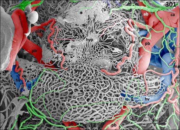
Pituitary gland. Vascular anatomy. Ventral view at the hypothalamo-hypophysial area. Shown is the arterial supply of the pituitary gland via terminal branches of the superficial infundibular artery (sia) and its drainage via the hypohysial vein (hv). Meningeal capillaries are colored green. ca cerebral carotid artery, dia deep infundibular artery, dl distal lobe of pituitary gland, hyp hypothalamus, il intermediate lobe of pituitary gland, me median eminence, nl neural lobe of pituitary gland, ab anterior branch of cerebral carotid artery, pb posterior branch of cerebral carotid artery. Asterisk marks large evasate.

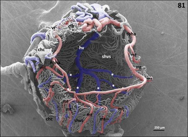
Superficial hyaloid vascular system of a left eye. Slightly oblique frontal view. Nasal is at left, temporal is at right. Two thirds of the chorio-capillaris (chc) and ciliary body (cib) are removed to expose the superficial hyaloid vascular system (shvs). Specimen is spatially distorted, but otherwise intact. Shown are nasal (nb) and temporal branch (tb) of the hyaloid artery (ha). Each semicircular branch gives off 7-10 meridional twigs (mt) which run towards the posterior pole of the eye. Further seen are the roots of the hyaloid vein (asterisks) between arterial meridional twigs (mt). hv hyaloid vein.
Fotos: Gefäßausgusspräparation / Vascular corrosion Casting (Quelle: COLOR ATLAS OF THE MICROVASCULATURE OF TISSUES AND ORGANS IN ADULT XENOPUS LAEVIS, A. Lametschwandtner und B. Minnich, in. prep.)


